


OCT Scan for Glaucoma: What It Can Show
Glaucoma often develops without pain, redness or a noticeable change in sight. By the time a person becomes aware of missing areas in their vision, damage to the optic nerve may already be established. That is why an OCT scan for glaucoma can be so valuable: it allows your optometrist to look closely at the structures affected by the condition, often before changes are obvious to you.
At Darlington Opticians, OCT imaging forms part of a thorough eye-health assessment where it is clinically appropriate. The scan is quick, non-contact and painless, but the information it provides can support earlier detection, careful monitoring and timely referral when needed.
What is an OCT scan for glaucoma?
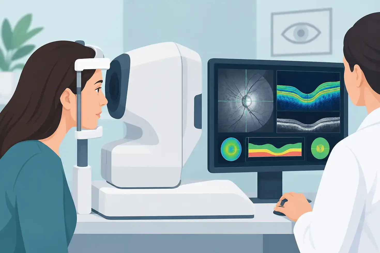
OCT stands for Optical Coherence Tomography. It is an advanced imaging test that uses light to create detailed cross-sectional pictures of the back of the eye. In glaucoma assessment, the main focus is usually the optic nerve and the nerve fibre layer around it.
The optic nerve carries visual information from your eye to your brain. In many forms of glaucoma, the delicate nerve fibres gradually become damaged. An OCT scan measures the thickness of these layers and compares the results with a reference database for people of a similar age.
This does not mean that a single red, amber or green result provides a diagnosis on its own. Eyes vary naturally, and scan findings must always be interpreted alongside the rest of your examination. However, OCT can reveal subtle structural changes that are not visible with standard examination alone.
What the scan looks at
During a glaucoma-focused OCT assessment, your optometrist may examine the retinal nerve fibre layer, which surrounds the optic nerve head, and the ganglion cell layer nearer the centre of the retina. These are areas where thinning can be associated with glaucoma.
The scan also produces an image of the optic disc, sometimes called the optic nerve head. Your optometrist considers its shape, size and appearance, as well as whether there are differences between your two eyes. The detail helps build a more reliable picture than any one test can provide.
Why glaucoma needs more than an eye-pressure check
Many people associate glaucoma with raised eye pressure. Pressure inside the eye is an important risk factor, and measuring it remains a key part of an eye examination. Yet pressure alone cannot confirm or rule out glaucoma.
Some people have higher-than-average eye pressure for years without developing optic nerve damage. Others develop glaucoma despite readings that fall within the usual range. This is known as normal-tension glaucoma. For that reason, a proper assessment considers several findings together: eye pressure, optic nerve appearance, OCT images, visual field results, family history and other personal risk factors.
An OCT scan is particularly useful because it assesses structure, while a visual field test checks function - whether glaucoma may be affecting what you can see. Structural change can sometimes be detected before a measurable visual field defect develops. Equally, a scan may look stable while other test results require further investigation. The value comes from combining the evidence and comparing it over time.
What happens during the scan?
The OCT scan itself usually takes only a few minutes. You will be asked to sit at the instrument and rest your chin and forehead on supports. Looking at a fixation target, you keep your eyes as still as comfortably possible while the scanner captures images. Nothing touches your eye and no radiation is used.
Your pupils do not always need to be dilated, although drops may occasionally be recommended if a clearer view of the back of the eye is needed. These drops can temporarily blur close vision and increase light sensitivity, so it is sensible to ask whether you will need someone else to drive you home.
A technician or optometrist will check that the image quality is good enough to interpret. Dry eye, blinking, a cataract, a small pupil or simply difficulty holding the eye steady can affect a scan. If an image is unclear, repeating it is normal and does not mean there is necessarily a problem.
Who may benefit from glaucoma OCT monitoring?
OCT is not only for people with diagnosed glaucoma. It may be recommended where there is a reason to look more closely at the optic nerve or establish a useful baseline for future comparison.
This may include people with a close relative who has glaucoma, raised eye pressure, an unusual-looking optic nerve, a previous suspect result, or visual field findings that need clarification. Risk also increases with age, and some medical conditions, medicines and eye conditions can influence the clinical picture.
If you have been referred to hospital eye services or are under shared care for glaucoma, repeat OCT imaging can help show whether the nerve fibre layers appear stable. Monitoring matters because glaucoma changes are usually gradual. A sequence of well-quality scans taken over months or years is often more informative than one isolated result.
Understanding your result without unnecessary worry
It is understandable to feel concerned if your optometrist mentions thinning, asymmetry or a result outside the normal reference range. These terms do not automatically mean you have glaucoma. The reference database cannot account for every healthy variation in eye shape, prescription or optic nerve size.
For example, people who are very short-sighted may have optic nerves that are more difficult to assess using standard comparison data. A cataract or poor scan signal can also influence measurements. Your clinician will consider whether the scan matches what they can see when examining your eyes and whether it agrees with pressure readings and visual field tests.
If the findings suggest possible glaucoma or progression, you may be referred for further assessment. This is a precaution designed to protect your sight, not a final diagnosis. Hospital specialists may repeat tests, measure pressure at different times, assess the drainage angle of the eye and decide whether observation, treatment or ongoing review is appropriate.
How often should an OCT scan be repeated?
There is no single timetable that suits everyone. If your eyes are healthy and there are no glaucoma risk factors, OCT may be used only when your optometrist sees a clinical reason. If you are considered a glaucoma suspect, have raised eye pressure or already have glaucoma, follow-up intervals depend on your individual level of risk and whether results are stable.
A repeat scan is most useful when it can be compared with earlier images taken using the same or a comparable method. This is one reason regular eye examinations at a practice that keeps a clear clinical record can be reassuring. It allows small changes to be assessed in context rather than relying on memory or a single appointment.
Do not wait for symptoms if you have been advised to attend for monitoring. Early glaucoma often causes no warning signs, while prompt treatment can help slow further damage where treatment is needed.
OCT is detailed, but it is one part of your care
OCT technology has strengthened the way optometrists assess glaucoma risk, providing precise images that would not be possible with an ordinary sight test alone. It is not, however, a replacement for the wider examination or for specialist assessment where this is required.
The most useful outcome is not simply receiving a scan report. It is having the result explained clearly, understanding whether any follow-up is recommended, and knowing who to contact if you notice a sudden change in your vision. Keeping regular appointments gives your eye-care team the best opportunity to spot meaningful change early and help safeguard your sight for the years ahead.
Comments